
ALCOR Life Extension Foundation. The World's Leader in Cryonics
Alcor.org | 2014
The Alcor Life Extension Foundation is the world leader in cryonics, cryonics research, and cryonics technology. Cryonics is the science of using ultra-cold temperature to preserve human life with the intent of restoring good health when technology becomes available to do so. Alcor is a non-profit organization located in Scottsdale, Arizona, founded in 1972.
The central vision for the Human Brain Project (HBP) - reconstructing and simulating the human brain - was developed by Henry Markram, based on the research strategy developed in the Blue Brain Project. Starting in 2010, Markram created and coordinated the consortium of 80 European and International partners that developed the original HBP project proposal. In January 2013, after multiple rounds of peer review, the project was selected as one of two FET Flagships, to be funded with 1 billion euro over 10 years. The project began operations in October of the same year. The project currently includes 112 partners.
 |
| Source: http://alcor.org/ |
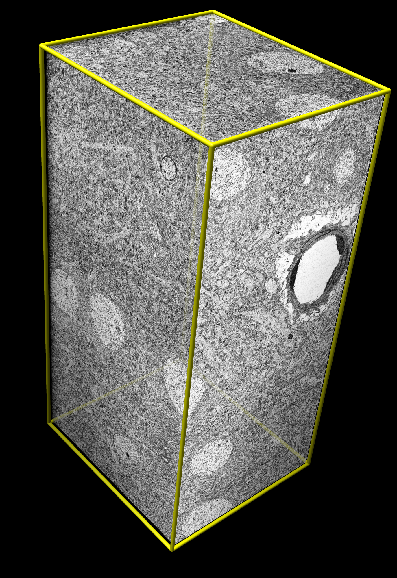 |
| Source: https://www.cgl.ucsf.edu/chimera/animations/ratbrain/ratbrain.html |
<more at http://alcor.org/; realted articles and links: http://bluebrain.epfl.ch/page-52741-en.html (The Blue Brain Project website) and http://www.ncbi.nlm.nih.gov/pmc/articles/PMC3716574/ (Large-Volume Reconstruction of Brain Tissue from High-Resolution Serial Section Images Acquired by SEM-Based Scanning Transmission Electron Microscopy. Masaaki Kuwajima, John M. Mendenhall, and Kristen M. Harris. Methods Mol Biol. 2013; 950: 253–273. doi: 10.1007/978-1-62703-137-0_15. [Abstract: With recent improvements in instrumentation and computational tools, serial section electron microscopy has become increasingly straightforward. A new method for imaging ultrathin serial sections is developed based on a field emission scanning electron microscope fitted with a transmitted electron detector. This method is capable of automatically acquiring high-resolution serial images with a large field size and very little optical and physical distortions. In this chapter, we describe the procedures leading to the generation and analyses of a large-volume stack of high-resolution images (64 μm × 64 μm × 10 μm, or larger, at 2 nm pixel size), including how to obtain large-area serial sections of uniform thickness from well-preserved brain tissue that is rapidly perfusion-fixed with mixed aldehydes, processed with a microwave-enhanced method, and embedded into epoxy resin.])>
No comments:
Post a Comment